As the duck flies: Avian influenza virus and migratory mallards

For a pathogen to survive it has to find new hosts to infect. This may sound simple, but if you consider the entangled mesh that is the biology of a host species you realize that there are plenty of ways that things can go wrong, stopping the chain of transmission. First of all, the harm the pathogen incurs on its host – the virulence – needs to be balanced between being too low – the infection will be cleared before any transmission opportunities have occurred –  or too high, so that it causes the demise of the host before transmission can take place. Second, the pathogens must overcome the hurdles of moving from one host to the next, be it in water, air or through the bite of an arthropod vector. And third, it has to overcome the fact that most hosts are not sedentary, but move varying distances in response to changes in the environment they inhabit. Finally, it needs to be able infect the new host and evade the immune system to establish infection. Not an easy feat, but something that is happening all the time in the world of viruses, bacteria, fungi, parasites and their hosts.

In the avian influenza field, the realization of the importance of bird migration in the epidemiology has a long history but we haven’t really been able to address it in the required detail. Most studies have addressed the process at the population level, inferring movements either from ring recoveries or from virus phylogenetic perspectives. If you have followed what we do, it will not come as a surprise that we are interested in both influenza viruses and bird migration. A longstanding goal for us has been to integrate virology and movement ecology to better understand the epidemiology of avian pathogens. This is where it gets exciting, as the technology needed for these types of studies are available. Last year we deployed loggers on migrating mallards at our main study site, the Ottenby Bird Observatory on the island of Öland in SE Sweden, and followed them during migration as a part of the H2020 program DELTA-FLU.

We programmed the loggers to record GPS-positions in bursts, hoping to retrieve as much data as possible during active flight. From the flight data we extracted the metrics of flight: how does a mallard migrate – how fast, how high, and in which direction? And how do these parameters change during the flight? These metrics formed the basis for a Mallard Migration Simulator with which we could simulate different types of migrations, based on the normal flight behaviors of mallards.

The next step was to use the ring recoveries retrieved from the study site over the last 50 years to get realistic headings of migratory flights. Finally, we introduced individual-level epidemiological parameters from our study populations and built classical SIR-models. Combined, this allowed us to look at the likelihood that a bird that migrated was infected with low-pathogenic avian influenza, and that it maintained infection during migration, controlled by season, age of birds and other factors that could contribute. The resulting data can be transformed into a risk map for transmission.

I am very pleased with this approach, and think it is a novel way of analyzing this type of data. The next step, of course, is to consider such models for highly-pathogenic avian influenza viruses on larger spatial scales. We are collecting tracks of four species of ducks in different parts of Eurasia and hopefully we will be able to make realistic models of virus dissemination among migratory ducks in a flyway perspective.

Link to the paper:

van Toor, M.L., Avril, A., Wu, G., Holan, S.H. & Waldenström, J. 2018. As the duck flies – estimating the dispersal of low-pathogenic avian influenza viruses by migrating mallards. Frontiers in Ecology and Evolution 6:208. doi: 10.3389/fevo.2018.00208

Flu transmission – a review

Hi there everyone, it is time for ducks and flu again! Just the other week we published a review on how host and virus traits affect transmission of low-pathogenic avian influenza viruses in wild birds. You should check it out – it is freely available in Current Opinion in Virology.

In this piece, Jacintha van Dijk, Josanne Verhagen, Michelle Wille and I synthesized the current knowledge of wild bird/flu interactions focusing on exposure and susceptibility. It is always challenging to write a review, especially when there are restrictions on length. But it is also fun (especially with such a talented team). We identified nine key host traits that can affect transmission: migration, non-migratory movements (e.g. dispersal), foraging, molt, reproduction, age, sex, pre-existing immunity and body condition, and provided the most recent findings from the literature regarding these traits. We also looked at five virus traits that can affect LPIAV transmission: virus stability, virus binding, virus replication, and the ability of the virus to evade the host innate and adaptive immune response. Many of these traits are not mutually exclusive, some have inherent spatial and temporal variation and can be affected by other confounding or unidentified factors.

Compared to many other wildlife pathogens, there is actually quite a lot of studies on LPIAV disease ecology to draw from. Yet, there is a clear need for additional and more integrative studies. You could say there are two sides: one more traditional approach with controlled infection experiment, and one more ecological approach with field samples and observations. Both are good, but neither is perfect. In lab studies, there is uncertainty in how well the experiment mimics the natural situation, and in field studies there are often many uncontrolled factors or results are correlative. We argue that these lines of research should be combined more often, either to use field studies to generate hypotheses to test in the lab with higher ecological realism, or to do semi-natural approaches in the field. A particularly challenging part is to study virus in free-living wild birds. Hopefully, the ongoing developments of remote-tracking could also be used to follow individual birds in the field for assessments of movement ecology, contact rates and other parameters of importance for predicting LPIAV maintenance.

Anyway, since the article is open access I strongly recommend you to click on this link and download the full review.

van Dijk, JGB., Verhagen, JH, Wille, M. & Waldenström, J. 2017. Host and virus ecology as determinants of influensa A virus transmission in wild birds. Current Opinion in Virology 28: 26-36.

The conundrum of influenza A virus diversity and host immune responses – lessons from a vaccination experiment in Mallards

The influenza A virus is in an interesting virus. It exists in many subtypes and can infect a range of hosts, but most of the variation in subtypes and lineages is restricted to wild waterfowl, especially dabbling ducks. Contrary to humans and other mammals, the virus doesn’t normally cause disease in ducks and these viruses are said to be low-pathogenic. The traditional explanation for the evolution of subtypes is that they have evolved to be sufficiently antigenically different that infection with one subtype does not incur protection to another one. Hence, antibodies raised against a H1 virus would do poorly with an H7 infection, and vice versa, but work well against an infection with a homologous virus, i.e. another H1 virus.

The latter is called homosubtypic immunity, and has been shown in a range of studies of Mallards (our favorite bird), using both experimental infections and studies conducted in the field, and although serum antibodies in Mallards seem to wane with time, immunity does seem to be long-lasting (see for instance Tolf et al. 2013).

A few years ago, we identified the existence of heterosubtypic immunity in wild Mallards. We analyzed infection histories of individuals recaptured during their stopover stay at Ottenby and investigated patterns of subtype occurrence compared to what would be if infection order was non-structured. In essence, what we could see was that heterosubtypic immunity was frequent, most strongly observed at hemagglutinin (HA) clade level, but also detectable at the HA group level. In contrast, there was no effect of the neuraminidase subtype (see Latorre-Margalef et al. 2013). The strength of this pattern was rather surprising, and has sparked follow-up studies.

Lately, a number of studies have used experimental infections to investigate heterosubtypic immunity further, either as a cause of understanding how highly-pathogenic viruses can be maintained in waterfowl, or for assessing immunity patterns in low-pathogenic avian influenza infections. Two nice, recent articles are by Segovia et al. 2017 investigating H3N8, H4N6, H10N7 and H14N5 infections in a balanced design, and by Latorre-Margalef et al. 2017 assessing protection of H3 antibodies against a range of other virus subtypes. Collectively, these studies suggest that the order of infections are important for future disease dynamics, both at the individual level but also at the population level. In other words: the order of outbreaks in a population will govern the fate of other subtypes in the population later; a competition among subtypes over susceptible hosts. This is very interesting, and something we currently try to model with infection history data of captured and recaptured wild Mallards at our study site.

The principle of immunity is that previous infections will render the bird immunity to reinfection with the same virus subtype, so called homosubtypic immunity, as long as the antigenic properties of the two strains are similar. A heterosubtypic immunity is when infection with one subtype provides full or partial protection against other subtypes, and it is expected that this is more common in phylogenetically related subtypes. (Illustration by M. Wille)

However, field and lab are two different things, and a couple of years ago we wanted to use the duck trap at Ottenby to study immune processes. As we cannot infect and release birds in the trap we used vaccination as a means of simulating previous infection. We prepared two vaccines, one against H3 and one against H6 (and one sham), immunized birds and followed them to make sure they developed serum antibodies (against NP) and neutralizing antibodies against the HA, after which we released them into the duck trap and followed their natural infections in the wild. As often is the case, our experiment didn’t really go as intended. First of all, there were no H6 infections in the wild population at the time of the experiment, thus no H6 infections recorded in any of the groups of our experiment so we couldn’t analyze the protectiveness of H6 vaccination. Quite surprisingly, all three groups were infected with H3 viruses – including the group that had received the H3 vaccine.

There are two possible explanations for the failed homosubtypic response. One is that immunization didn’t result in protective immunity, and the other that the viruses were antigenically different. We did detect neutralizing antibodies against H3 viruses in the ducks, suggesting these ducks did raise a specific immune response against the vaccine. Interestingly, the ducks didn’t raise a similar response against H3 infections after being in the duck trap. Investigating the latter we could show that the vaccine strain and the outbreak strain differed by a number of substitutions close to the receptor binding site. Going back to our virus neutrilizations, we could see differences in in the strength of the antibody response against different H3 viruses, including differences between the strain we used to vaccinate and the strain that was circulating during our experiment. Sufficiently different to suggest antigenic difference. The paper is just out (Wille et al. 2017). H3s are quite interesting, as they have been the focus in much of human infection research, especially because there seems to be two antigentically different lineages and after infection with one of these H3 lineages humans may not be protected against the other. Antigenic cartography has identified the importance of a few sites in or at the receptor binding site for immune evasion in human H3N2, and it is possible that this is what we see also in avian H3s.

A protein structure of the H3 hemagglutinin, where differences between the outbreak and the vaccine strains are mapped. For more information have a look at the paper in Molecular Ecology.

So, what can we learn from this? As always in science, each new study answers some questions but raises many more. First of all, what is the rate of antigenic drift in avian viruses, how do that differ among subtypes, and what does that mean in a functional and evolutionary context? How does this relate to long-term subtype dynamics and the role of herd immunity and heterosubtypic immunity in wild avian hosts? Second, it illustrates our lack of knowledge on the actual mechanisms of immunity –  despite low-pathogenic avian influenza viruses being gastrointestinal infections in waterfowl, we tend to study serum antibodies rather than mucosal antibodies or innate immune responses. Third, we have work to do as regards vaccination as a model for disease – are immune processes the same, and is protection similar?

Stay tuned – we will get back to this subject later.

If you want to read the study, it is available as Open Access:

Wille, M., Latorre-Margalef, N., Tolf, C., Stallknecht, D.E. & Waldenström, J. 2017. No evidence for homosubtypic immunity of influenza H3 in Mallards following vaccination in a natural experimental system. Molecular Ecology. [doi:10.1111/mec.13967]

http://onlinelibrary.wiley.com/doi/10.1111/mec.13967/full


Seabirds and flu, a review

A small murre colony on Cabot Island, Canada.

[This post is by Michelle Wille, postdoctoral researcher at Uppsala University]

For those who have visited a seabird colony, you would know that it is a loud and crowded place, with large swaths of the colony covered in guano. It literally stinks of bird poo. If you were to imagine a good host for a virus that is transmitted by the fecal oral route, one could imagine that these conditions would be excellent for transmission. A virus, such as the influenza A virus (IAV).


This virus is one of the most important and well-studied avian viruses, especially in its reservoir hosts, the dabbling ducks. However, for seabirds – the majestic creatures that roam the oceans – no real synthesis has been published despite close to 50 years of surveillance. In fact, when I started working on IAV in seabirds, we knew very little about the presence and prevalence of influenza in this group of birds. What we did know was that seabirds were being sampled for influenza – in fact, most bird groups were being sampled for IAV following the highly pathogenic H5N1 outbreaks after 2005 – but we didn’t actually know how seabirds fit into the ecology of influenza. Are they infected? Are some seabirds more important than others? Do they follow similar patterns to ducks or gulls? Are their viruses unique, or more similar to duck or gulls?


Antarcric Tern

Antarctic tern

We set out to collate the existing knowledge on IAV in seabirds – a diverse collection of species and are best defined through their shared propensity to spend portions of their lives at sea – and pulled together as much surveillance data as possible from publications and influenza databases to try to evaluate sampling effort in seabirds, and which species play a role in IAV ecology. This review was just published in the journal Avian Diseases. It turns out, scientists have sampled a large number of seabirds over the last 50 years: 41,828 samples from 98 species, spanning 14 avian families in 6 orders. This may seem like a lot of samples, but if broken down it equals only 8.5 samples per species per year. To put it in perspective, from our sampling site in Sweden, 22,229 samples were collected from Mallards between 2002-2009, and it is samples sizes like these that allow us to make stronger inferences on IAV ecology.


While this illustrates the lack of effort overall, some seabirds have received more effort and attention. Terns as a group are heavily sampled, although sporadically rather than systematically. Terns are interesting as the first confirmed outbreak of highly pathogenic influenza in wild birds occurred in Common Terns (Sterna hirundo) in South Africa back in 1961. Despite very few isolations of viruses, serology suggests circulation of IAV in terns and noddies and a diversity of virus subtypes – most recently highlighted in the Indian Ocean system. Most interesting, perhaps is the compelling evidence suggesting that Murres/Guillemots (Uria sp.) are hosts for IAV. Research to investigate IAV in murres dates back to the 1970s, and interest in these birds has been renewed with increased sampling effort in the past 10 years. These birds are piscivorous, limited to the northern Holoarctic where they breed predominantly on islands, often on steep cliffs. Within all the seabird groups, the greatest number and diversity of viruses come from murres, with viruses isolated across their range – Russia, Sweden, Greenland, Newfoundland (Canada), Nunavut (Canada), Alaska (USA), and Oregon (USA). Unfortunately there is rather limited serological information in Common and Thick-billed Murre, which would provide a more long-term assessment of influenza dynamics.


COMU

A few other species/groups have large enough sample sizes to estimate IAV prevalence with confidence, but serology, despite small sample sizes, indicates IAV presence in most seabird species tested. However, more focused work is required to better assess these species as hosts. Regardless, if you are interested in the IAV status of the seabirds you work on – sampling effort and IAV results are presented for all 98 species.


concept

What is the role of seabirds in the epidemiology of low-pathogenic avian influenza?

What was a surprise for us, as we were completing this review, was how little we could say about the role of seabirds in the ecology of seabirds due to limitations in sampling. There is clearly a space to fill for an aspiring IAV researcher. If you want to sample for IAV and be able to draw some conclusions – here are some things to think about:


  1. Influenza A in birds is seasonal. Some months the prevalence is high (up to 30%) and some months it is low (>0.00001%). While seabirds are logistically hard to access, temporal and repeated sampling is key.


  1. Within an individual, the period of shedding live virus is very short. While longer periods have been detected (up to 14 days), usually birds shed viruses for less than 7 days. This highlights the importance of serology, or assessing the antibody prevalence in a population. This allows us to ascertain whether the population has been infected by IAV in the past, and therefore, whether it is a population to target (if positive).


  1. Seabird colonies may have many species, and it is tempting to take a few samples from each species present. Low sample size however limits the detection probability. For example, if prevalence of IAV is about 1% in the population, you need to take well over 100 samples to have a 95% probability of detecting the virus. Putative prevalence of IAV in seabirds is in this 1% range.


  1. Maintaining “cold chain” is key. Seabird colonies are logistically hard to sample, and dragging a -80C freezer or vapour shipper may just not seem to be worth the effort. But, RNA viruses degrade rather rapidly, and swaths of negative samples may be false negatives due to poor sampling handling. While it is speculation, perhaps the reason that we are starting to be more successful at isolating influenza from Antarctic Penguins is an improvement in cold chain (who would have through it would be difficult to keep samples at a constant temperature of -80C in Antarctic!).


I feel privileged to be writing this piece after recently spending a week working in a Murre colony in Sweden. Seabird colonies really are the best places to be – serene beauty on the steep, the smell of guano-ladened cliffs on (remote) islands, with the flutter of murre wings and peeping of recently hatched murre chicks.

Link to the article:

Andrew S. Lang, A.S., Lebarbenchon, C., Ramey, A.M., Robertson, G.J., Waldenström, J.& Wille, M. 2016. Assessing the Role of Seabirds in the Ecology of Influenza A Viruses. Avian Diseases 60(1s):378-386.


Penguins

Adelie and Gentoo penguins doing their thing.

Influenza A virus epidemiology – from individual disease histories to disease dynamics

5566951033_902c131ed8_b

Mallards on the wing (Photo by Flickr user Bengt Nyman used under a CC-BY 2.0 license)

Wildlife disease studies are challenging. That’s a fact. If you want an easy science life you should choose another path with more instant results. However, challenging is also the opposite of boring, and the rewards of getting your results are even more exhilarating when lots of toil, sweat and tears have been invested. As readers of this blog are aware, wildlife disease studies are what we do, and I have repeatedly written about our ongoing work on influenza A virus ecology and epidemiology in wild migratory Mallards. This week another study from our study site was published, entitled Capturing individual-level parameters of influenza A virus dynamics in wild ducks using multistate models, which can be found on early view in the Journal of Applied Ecology.

The challenges of studying wildlife disease dynamics are that you want to capture a dynamic process influenced both by the host and the pathogen, which in turn is compounded by variation in the environment – both biotic factors, such as food abundance and the occurrence of other potential hosts, and abiotic factors, such as weather and climate. Disentangling these interconnected effects is a little like making a cube out of mercury. In most wildlife disease studies the available data is at the population level, usually in the form of prevalence rates at specific time points. This type of data is ‘fairly easy’ to collect – you head out into the field, sample all animals you can lay your hands on and then use this snapshot in time as a proxy for the true disease dynamic in your system. The more times you are out collecting data, the better your model becomes. However, disease is driven by factors operating at the level of individuals, such as infection risk and recovery rate, and that type of data can only be acquired by repeated sampling of individuals across a suitable timescale. This is rarely achieved because of logistical, practical and monetary reasons.

26383076326_62d7d29903_k

Mallards on the wing (Photo by Flickr user Bengt Nyman used under a CC-BY 2.0 license)

We, however, sit on a huge collection of Mallard and flu data gathered at the same study site with similar methods over a period of close to 15 years. Our latest paper, headed by Alexis Avril and with collaboration with colleagues in France, utilizes this dataset to develop individual-based influenza A virus epidemiological models. This proved to a monumental task that stretched over several years and burned the processors of a good number of computers. Part of the difficulty can be attributed to the data itself – capture and disease histories for 3500 individuals collected over 7 seasons, where at each capture occasion axillary data on bird age, sex, condition, infection status and weather were included. But also the patchy nature of recapture probability and the short duration of most influenza virus infections contributed significantly to extensive data crunching.

Picture2

The conceptual framework in the multistate CMR model.

The method we used was multistate capture-mark-recapture models, which are extensions of models originally developed to investigate mortality rates from census data, but where one can include the infection state – i.e. infected or not with influenza virus – as a factor in the analyses. Interested readers should head over and read the publication, as I will spear the rest of you any hardcore statistics and model lingo. Parts of the abstract serves as a good summary:

 For most years, prevalence and risk of influenza A virus (IAV) infection peaked at a single time during the autumn migration season, but the timing, shape and intensity of the infection curve showed strong annual heterogeneity. In contrast, the seasonal pattern of recovery rate only varied in intensity across years. Adults and juveniles displayed similar seasonal patterns of infection and recovery each year. However, compared to adults, juveniles experienced twice the risk of becoming infected, whereas recovery rates were similar across age categories. Finally, we did not find evidence that infection influenced the timing of emigration from the stopover site.

Our study provides robust empirical estimates of epidemiological parameters for predicting IAV dynamics. However, the strong annual variation in infection curves makes forecasting difficult. Prevalence data can provide reliable surveillance indicators as long as they catch the variation in infection risk. However, individual-based monitoring of infection is required to verify this assumption in areas where surveillance occurs. In this context, monitoring of captive sentinel birds kept in close contact with wild birds is useful. The fact that infection does not impact the timing of migration underpins the potential for mallards to spread viruses rapidly over large geographical scales.

Our findings corroborate much of the earlier works done on IAV in birds from population level data or from infection experiments, but with higher robustness of the conclusions. Importantly, we provide estimates of the most crucial infection parameters and show how they vary in relation to age in different seasons and years. And from a model point of view, we show that MS-CMRs are a potent method for disease dynamic inferences. We hope this paper will be read and cited by people in the IAV field and in general disease dynamic research, and that it will be useful for stakeholders interested in the contribution of wild birds in the epidemiology of IAV in poultry.

Link to the paper:

Avril, A., Grosbois, V., Latorre-Margalef, N., Gaidet, N., Tolf, C., Olsen, B. & Waldenström, J. 2016. Capturing individual-level parameters of influenza A virus dynamics in wild ducks using multistate models. Journal of Applied Ecology, online early.

On the cover

In the Swedish academic system a PhD thesis is an actual printed book. Yes, a proper book-looking book. A kind of book you can shove in the hands of your family and say ‘here it is, the fruits of my labor, the sum of all my forsaken years, MY THESIS!’

The editions are small, usually between 100 and 200 copies, of which 30 go to specified libraries, kept safe in the hallways of science. One is nailed to an oak tree three weeks before the thesis defense, thereby symbolically presented to the public (and continuing a tradition dating back to Martin Luther’s nailed thesis to the church port of Wittenberg in 1517). Although it probably isn’t really necessary to print them on paper in this age of computers, it is a tradition worth honoring. More than anything it represents a closure of sorts, and a mark of something new.

I really like that feeling when the book gets back from the printers and the student finally sees the end of the wonderful but torturous path that is a PhD. The making of the book is sometimes also an act of procrastination, where the student can spend an inordinate time adjusting the colors on the cover, rather than finalizing that final chapter.

Below are the theses produced in our research group in Kalmar the last years. If you want to read them there are links to the online publications too.

Daniel Bengtsson

• Daniel Bengtsson, Linnaeus University, Stopover ecology of mallards – where, when and how to do what? 11 March 11 2015, ISBN 978-91-88357-00-7.

• Michelle Wille, Linnaeus University, Viruses on the wing: evolution and dynamics of influenza A virus in the Mallard reservoir, 8 May 2015. ISBN 978-91-87925-56-6.

Johan Stedt

• Johan Stedt, Linnaeus University, Wild birds as carriers of antibiotic resistant E. coli and Extended-Spectrum Beta-Lactamases, 13 June 2014. ISBN: 978-91-87427-93-0.

Petra Griekspoor Berglund

• Petra Griekspoor, Linnaeus University, Exploring the epidemiology and population structure of Campylobacter jejuni in humans, broilers and wild birds, 28 May 2013. ISBN 978-91-87427-83-1.

Neus Latorre-Maraglef

• Neus Latorre-Margalef, Linnaeus University, Ecology and epidemiology of influenza A virus in Mallards Anas platyrhynchos, 8 June 2012. ISBN 978-91-86983-61-1.

Jorge Hernandez

• Jorge Hernandez, Uppsala University, Human pathogens and antibiotic resistant bacteria in the Polar regions, 10 October 2014. ISBN 978-91-554-9016-4.

Jenny Olofssons avhandling

Jenny Olofsson, Uppsala University, Amoebae as hosts and vectors for spread of Campylobacter jejuni, 2015. ISBN: 978-91-554-9276-2.

Flu, ducks and the costs of being infected

IMG_4978

There was light snow this morning, but it has since melted away, leaving small puddles on the streets. Unfortunately, the sun seems to have lost today’s battle with the fog and the low clouds – it is, in essence, an ordinary wet, cold and gloomy February day. But if I peer out through the window, ignoring the construction works in the foreground, there is water on the horizon. And where there is water, there are ducks. And where there are ducks, there is flu. One cannot ask for more.

Over the years I have thought much about ducks and flu. (Some would say too much, but they don’t know what they miss). Although my research group has already produced four PhD theses on this topic, there is so much more that I would like to know. Some of it is  highly specialized knowledge, of interest for a limited set of like-minded scientists with  acquired duck disease tastes. Other things are quite basic, but hard to study, such as the question whether ducks infected with flu suffer from infection or not. That is a pretty important question also for a broader audience, as it has relevance for how well virus can spread with individuals in the environment; especially how ducks may spread virus long distances during migration. So, do they suffer from infections, or not?

Actually, there has been some controversy on this topic – partly stemming from different methods of quantifying disease effects. A field ecologist and a veterinarian have different scales in their toolboxes, one could say. In the latter case, disease signs are determined in  experimental infections in animal house facilities, where individuals can be followed over time. Such experiments in Mallards have not been associated with strong disease signs – as long as we consider the low-pathogenic avian influenza viruses that are naturally occurring in wild avian populations (highly pathogenic AI is a completely different story). Infected Mallards shed viruses, but are otherwise apparently healthy, or only display a very short increase in body temperature. But, the ecologist argues, the artificial environment with plenty of food, controlled temperature and absence of predators is not really mimicking the situation in the wild, where even small reductions in vigilance and movement capacity may end in the death from a raptor’s claw. Absence of overt disease is not equal to absence of ecological costs, the ecologist would conclude.

The field studies so far have been a mixed bag, ranging from large effects to negligible effects depending on study and the species considered. The largest effect was seen in a study of Bewick’s swans in the Netherlands, where infected birds had poorer condition and migrated slower than uninfected swans. Such large effects have not been seen in other species, and one can not conclusively rule out other underlying factors, as the swan study was based on a limited number of birds. When it comes to Mallards – the most glorious of all avian influenza reservoir species – previous population studies from our group have suggested infected birds to weigh on average less than uninfected birds at capture.

Averages and populations are all and well, but to get to a mechanistic understanding one is better off with experiment conducted on a set of individuals. However, a problem is that we can not infect birds and release them in the field; in fact, we are not allowed to do so – there is a reason infection experiments are conducted in biosafety labs, after all. What to do, then? Well, we approached this question via GPS and accelerometer loggers attached to two groups of birds caught during the ongoing surveillance at our study site: one group of 20 Mallards with natural avian influenza infection at the time of capture, and another group of 20 Mallards that were negative for influenza at the time of capture.

The benefit of these data loggers is that they record such a wealth of information. From the GPS fixes we can follow the birds in the landscape and quantify their movements at spatial and temporal scales; from the accelerometer we can get metrics that describe activity, defined as movements in the x, y, z-dimensions. We predicted that infection would significantly hamper movement, and that with time the difference between infected and uninfected birds would level off (see figure below); hence the analyses need to take time in to account, too.

F1.large

Theoretical predictions of the influence of infection on movement metrics. If infection affects spatial behaviour, infected (blue) and uninfected (red) birds should behave differently at the time of release. We postulate that, at this time, movement metrics for infected birds should be lower than for uninfected birds, which would be revealed as different intercepts of the regression of the movement metrics against time for uninfected (β0) and infected birds (β0+βInf). As infected birds recover with time, their movement metrics will approach and eventually meet the values for uninfected birds. This happens when the slope of the regression of the movement metrics against time for infected individuals (βT.aft.Rel*inf) reaches the slope for uninfected birds (βT.aft.Rel), which is expected to be null.

The full paper is freely accessible at Royal Society Open, and I hope readers with a more heavy interest in movement ecology download and read it. There is a lot of data crunching and statistics there that most of you are likely not that interested in – if you are, go read the original publication – but remember even easy questions may be hard to answer. Okay, with that said, what where the results?

Well. There were no effects of infection on the movement parameters measured, at all. Yes, there were differences among individuals, and between night and day, but infection status did not explain much of the variation in movement metrics. This means that under the natural situation in this study, conducted during stopover in autumn migration, infected ducks moved as much as uninfected ducks. This also means they likely are not impaired by infection during active migration, and could therefore carry LPAI viruses on the wing as they depart the stopover site.

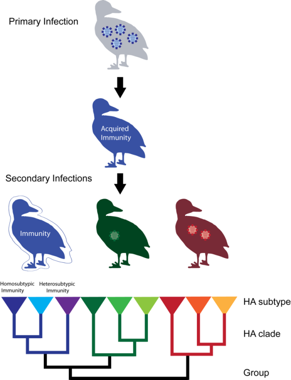
Is this, then, the last nail in the ‘cost of infection’ coffin for low-pathogenic influenza in ducks? Probably not, because one could argue that non all viruses behave the same (in fact, there should be a variation for virulence), and that some viruses may have adapted to infect non-mallard-birds, and hence be spillover infections in Mallards (and then potentially be at less than optimal virulence). Moreover – and perhaps a stronger argument – there may be differences in outcome depending on whether it is a primary infection, or subsequent infection; where the first infection in a naïve bird could be believed to carry a larger cost. Or there may be effects seen only at certain environmental conditions.

All these ‘but, or, perhaps, mayhaps’ are classic scientist disclaimers… My personal belief, these days, is that also the ecological costs of infection are slim. But I am happy to be proven wrong – out you go now and study.

There is water at the horizon still. And questions aplenty.


Link to the article:

Bengtsson, D., Safi, K., Avril, A., Fiedler, W., Wikelski, M., Gunnarsson, G., Elmberg, J., Tolf, C., Olsen, B. & Waldenström, J. 2016. Does influenza A virus infection affect movement behaviour during stopover in its wild reservoir host? Royal Society Open Science 3: 150633.
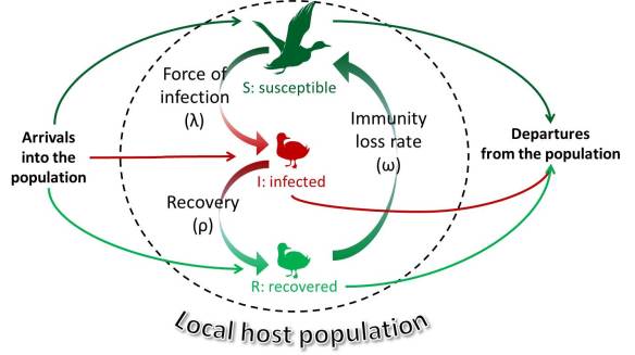
 

Older ducks poop fewer viruses – a story of how to sell a story (and some science, too)

Flickr user ‘John K’ under a CC BY-NC-ND 2.0 license.

Flying Mallards – great shot by Flickr user ‘John K’ (distributed under CC BY-NC-ND 2.0 license).

Last Friday, we published a paper in Applied and Environmental Microbiology with the title ‘How does sampling methodology influence molecular detection and isolation success in influenza A virus field studies?’ The study is a thorough analysis of 26,586 samples collected for detection of influenza A viruses from Mallards over some 9-10 years. The aim was to tap from the knowledge base gained in our long-term studies and provide some advice on best field and lab practices for others that want to initiate similar surveillance schemes for this virus. Thus, this is an article with high relevance for a limited crowd of people in our field, but not one that will attract hordes of interested ecologists. But as a methods paper it will be widely cited and useful.

In the paper we show which type of sample methodology that gives the best results. Not surprisingly, molecular detection was more sensitive than isolation, and virus isolation success was proportional to the amount of RNA copies in the sample – e.g. the more virus the easier to grow them in eggs. Comparing the results from specific RRT-PCRs and from isolation it was clear that co-infections were very common in the investigated birds. The effect of sample type and detection methods warrants some caution for interpretation of results of surveillance data, which we discuss in the paper.

Publishing a paper is great, but ideally the work doesn’t end there. One can blog about it, like I often do here, or one can try to push for the results in conventional media. The latter is tricky, and not something we do for every paper. However, this time we had some time to spare, and drafted a press release and sent it merrily along to the university’s Communications Office. One would think that a methods paper is a hard sell, but no – it was picked up by roughly 40 Swedish newspapers (mainly in short electronic form). That is pretty amazing! The reason? A catchy headline, of course.

The press release lifted up a smaller part of the paper, namely that for a given Ct-value, the isolation success was lower in samples from adult birds than from juveniles. This could be interpreted as adult birds (having had exposure to virus in previous infections) being more adept in limiting infections, manifested as less shed functional/infectious virus per given Ct-value. This is an interesting result that has bearing on our view of the epidemiology of the virus in the wild reservoir – but it was a side issue, not the main focus of the article. Anyway, during a sampling trip to Ottenby, we came up with the headline ‘Older ducks poop fewer viruses’ (Äldre änder bajsar färre virus). And this, my friends, is a title that was catchy enough to carry through the noise.

Communicating your science is important for a bunch of reasons, plus it is a part of the job description of us academics. But it is quite hard, and rewards at times unpredictable. Over the years, our research on ‘what scary deadly bugs do‘ has received much media attention. This is partly because the topic as such is appealing, but more likely that we actually put an effort into it. And time with media is time well spent. Anyway, that was a story about how to sell a story. For the real story please read the original article:

Latorre-Margalef, N., Avril, A., Tolf, C., Olsen, B. & Waldenström, J. 2015. How does sampling methodology influence molecular detection and isolation success in influenza A virus field studies? Applied and Environmental Microbiology, ahead of print, doi: 10.1128/AEM.03283-15.

With reference to reference genes – or, you’re doing it WRONG!

Allegorically, science is sometimes a meandering river. Photo by Flickr user Tim Haynes, used under a CC BY-NC-ND 2.0 license (And what a great shot it is!)

A lab is an organic thing that evolves over time. People come and go, grad students graduate and the research shifts; sometimes slowly like a meandering river, other times fast like a ricochet from a bullet. Three years ago our lab started a new project, aimed at understanding the role of innate immunity of ducks for fending off pathogens. Contrary to the adaptive branch of the immune system (T-cells and B-cells, antibodies and the like), the innate immune part is still mostly a black box for wildlife diseases. It contains a bunch of forces, from patrolling cells, eating bugs, to specialized molecules that stamp little holes in bacterial membranes or that ring the alarm bell to recruit other diverse armed forces. In ten years time ecologists will be all over the place with innate immunity – remember where you read it first!

I love to start new projects! Its great! Everything is possible! You get that pioneering feeling, chartering unknown areas for SCIENCE! Yay! However, after some time a realization dawns that there is a quite a long starting stretch before you can harvest. If you try to pick just the cherries, you run the risk of making uninformed choices, simply because there may be fundamental stuff specific to this new field that you were unaware of when you started. In our case, we are just about to publish some really cool stuff on mallard innate immunity, ranging from the evolution of innate immune genes to the actual responses of these genes upon infection. There is so much awesomness in that project that it could be the sparkle for a whole new lab. But before getting where we are at now, we have spent an eon in the lab optimizing and tinkering with protocols. You see, it is all about the little things, as finding the right primers, or the best way of extracting RNA from tissue samples.

A little while ago, we published a study that was part of this learning curve. What we wanted to do was to look at upregulation of a particular part of the innate immune system. We had done an infection experiment on mallards, testing whether natural infection with influenza A virus initiated increased expression of some target genes in the host. However, in order to be able to really say that a gene is up- or down-regulated, you need to make sure that you normalize your data to genes with stable expression, i.e. genes that do not respond in specific ways to infection.

Doing that exercise, we found that most of the studies previously conducted in our field had used genes that weren’t really stable. This prompted us to take a broader look at the literature to see how things are done, relative to how it should be done. This meta-analysis was published in PLOS ONE two weeks ago.

In short, we show that despite a common approved methodology, researchers still use too few reference genes and in most cases do not make sure that these genes really are stable in their study organism, in the tissue of choice, and under the experimental system under study. Or to quote parts of the abstract:

Recent guidelines have specified that a minimum of two validated reference genes should be used for normalisation. However, a quantitative review of the literature showed that the average number of reference genes used across all studies was 1.2. Thus, the vast majority of studies continue to use a single gene, with β-actin (ACTB) and/or glyceraldehyde 3-phosphate dehydrogenase (GAPDH) being commonly selected in studies of vertebrate gene expression. Few studies (15%) tested a panel of potential reference genes for stability of expression before using them to normalise data. Amongst studies specifically testing reference gene stability, few found ACTB or GAPDH to be optimal, whereby these genes were significantly less likely to be chosen when larger panels of potential reference genes were screened. Fewer reference genes were tested for stability in non-model organisms, presumably owing to a dearth of available primers in less well characterised species.

Another way of phrasing it would be: You’re doing it WRONG!

Link to the article:

Chapman, J. & Waldenström, J. 2015. With reference to reference genes: a systematic review of endogenous controls in gene expression studies. PLoS ONE 10(11): e0141853. doi:10.1371/journal.pone.0141853

Do wild birds give you campylobacteriosis?

Blackbird, Turdus merula. Photo from Flickr under a CC BY-NC-ND 2.0 license.

Blackbird, Turdus merula. Photo from Flickr under a CC BY-NC-ND 2.0 license.

There is magic in large numbers. Most often we scientist – regardless if we are field scientists or lab rats – struggle with acquiring sufficiently large sample sizes for the statistical tests we have set out to do. There are ways to deal with sparse data, but nothing beats a good-looking huge dataset if you want to test your hypothesis with confidence. Moreover, given that every biological system we measure has a degree of uncertainty, so called noise, means that if we are to find effects that are small we need to collect a lot of data.

Earlier this year, I co-authored a publication on Campylobacter epidemiology that really took advantage of large numbers. In this case, Cody et al. investigated if people get campylobacters from wild birds. This is something that has been suspected given the huge impact domestic poultry has – the single largest source of human campylobacteriosis – but not really proven. Over the years, the lab in Oxford has collected an enormous  dataset on the occurrence of Campylobacter jejuni in patients in Oxfordshire, UK. Not only is there a lot of data, each and every clinical case is associated with a genotyped bacterial isolate. That is an awesome treasure trove to investigate.

In this study, 5628 genotyped clinical isolates from Oxfordshire were run in a STRUCTURE analysis to try to associate each isolate with a putative source. The rationale here is that there are distinct sets of C. jejuni genotypes in different types of animals, especially in different species of birds. And as campylobacteriosis is a zoonotic infection with little to non human-to-human transmission such an analysis can indicate the degree of relevance of different sources for human epidemiology.

Did that sound awfully advanced? Perhaps. It really is quite simple. Consider you make a row of bins. Each bin gets a name, such as ‘chicken’, ‘cattle’, ‘goose’, ‘blackbird’ etc. Then you take each bacterial isolate in your hand, scrutinize it and put in a bin that you think it fits best in. A little bit like a sorting box for children. Starshaped objects go into the starshape hole, square objects in the square hole, etc. Except that it in this case it is the degree of resemblance at the genetic level that decides whether an isolate should be grouped with a particular source. The second thing is that you let the computer rerun this procedure over and over again until you get a probabilistic assignment to each bin.

STRUCTURE

The principle of STRUCTURE analysis.

In this paper, it was shown that the proportion of clinical isolates from Oxfordshire attributed to wild birds was 2.1%-3.5% each year. That is way lower than the values for chicken products, but given the very high incidence of campylobacteriosis in the human population it still means a large number of actual infections caused by bacteria that normally are found in wild birds. Which wild birds, you may ask. Primarily thrushes, is the answer – at least in Oxfordshire. The blackbird and the song thrush are two common garden birds that like to live close to us humans. Looking at the seasonal variation, the analysis showed that wild bird associated campylobacteriosis cases was more common during the warmer months of the year. This makes sense, as it is in summer when we loiter around in our gardens, and in nature, eating fruits and vegetables potentially contaminated with bird feces.

There is magic in large numbers, for sure.

Link to the paper:

Cody, A.J., McCarthy, N.D., Bray, J.E., Wimalarathna, H.M.L., Colles, F.C., Jansen van Rensburg, M.J., Dingle, K.E., Waldenström, J. & Maiden, M.C.J. 2015. Wild bird-associated Campylobacter jejuni isolates are a consistent source of human disease, in Oxfordshire, United Kingdom. Environmental Microbiology Reports 7: 782-788.