The slow pandemic

By Jonas Waldenström

When I write these lines, the storm Simone is hitting southeast Sweden. The trees around our house seem to move erratically, trunks bend, and the few remaining leaves are shaking. Things twirl in the wind, and strange tweaking sounds are coming from the chimney. A night better spent indoors, safe and sound. Perhaps, like me, sipping on a nightcap, and pray you don’t wake up to a devastated neighborhood.

Apart from the storm – which the news says is THE WORST in ages (which they often say) – the rain and the compact darkness are hallmarks of the season. The transition from autumn to winter is concomitant with an increase in the prescription of antidepressants. People, each and own, sit in their cabins and vegetate and wait for the spring, and the return of life to our north latitudes. Most depressing to me is the farewell to birds. No more fluttering songbirds in the garden, no aerial insectivorous swifts, no foliage gleaners, just the odd wet, miserable Jackdaw hoping to get lucky with the garbage bin.
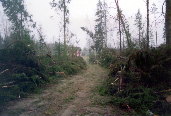
The aftermaths of Gudrun, the worst storm in recent times. It remains to see how the world looks tomorrow, after Simone.

The aftermaths of Gudrun, the worst storm in recent times. It remains to see how the world looks tomorrow, after Simone.

It is also the Halloween season, or the All Saints’ Day as it is remembered for here in Europe. A time when we should be scared by gauls and ghosts, real or in costumes. But do you know what scares me the most? Truly. It is not the (imminent) zombie apocalypse, nor is it motor bikers with baseball bats – no it is tiny little organisms. You might have heard their names before: MRSA, ESBL, or NDM1.

There is a silent pandemic out there. Not the influenza crash-and-burn style pandemic. No, a silent one, a slow step-by-step walk towards the abyss. In a not too distant future (actually already today) some pathogenic bacteria will be resistant to the antibiotic drugs we use to treat infections. Widespread resistance changes medicine, it changes the way our healthcare function, and the options a patient has for treatments. It changes the outcome of infection, it causes deaths. For instance, multi-resistant tuberculosis strains are spreading that can cope with almost all the compounds normally used for treatment.

We don’t have too look far to find resistant bacteria – a trip to your local hospital is sufficient. In that ecosystem, we select for bacteria that can withstand drug therapy – the results are nosocomial infections, or hospital-acquired infections. It has been estimated that a fifth of all infections are nosocomial, meaning that this is an acute and severe problem for the health sector. Thinking of it, actually, you don’t have to go to the hospital to find resistant bacteria. Simply opening the door to the fridge or the freezer will get you there. An increasing proportion of the food items we eat, especially meat, but also vegetables, contain bacteria that carry resistance genes.

A fridge. A clean fridge. But the bugs don't show.

A fridge. A clean fridge. But the bugs don’t show.

Bacteria have always evolved resistance to the chemicals used to fight them. We humans have waged war on bacteria for approximately a hundred years. Antibiotic compounds were an enormous success initially. Deadly infections were now made harmless and possible to control by prescription of drugs. When wound infections were treatable surgery became commonplace, and not a last resort. Not only appendicitis and cancers were treated, antibiotics opened the possibilities of today’s modern surgery where even organs can be moved between patients.

But what we didn’t think of in the early hay days of antibiotics was the vast evolutionary history of bacteria and chemical warfare. Although the human ingenuity is commendable, there is yet no compound invented that has not been tried in the evolutionary battle between microorganisms. Remember, fungi have tried to kill bacteria for billion of years. And through horizontal transmission between bacteria – sometimes across genera or families – genes evolved in one environment can find a home in a new bacterium and render the receiver new properties. A scientific niche the last few years have been to go searching for antibiotic resistance genes in preserved sediments, for instance in permafrost in Siberia, or in the deposits in caves or closed lakes. These studies have shown that, without a doubt, the genes have been around for a looooong time, thousands of years before humans even spoke the English language.

Actually a very poor representative of a scientific petri dish. In my lab that one likely was on its way to the autoclave. But you get it: bugs and gloves = dangerous

Actually a very poor representative of a scientific petri dish. In my lab that one likely was on its way to the autoclave. But you get it: bugs and gloves = dangerous

If you have read this far your either drunk or a masochist. My intention was to write about our latest article, exploring the occurrence of antibiotic resistance in wildlife in Chile, but the storm brought on the doom and gloom feelings. We’ll talk about solutions another night, when the storm winds are not howling across the land. But remember to be afraid – very afraid – of the slow pandemic that is about to change our way of life, as we know it.

be_afraid

Smell the roses –how does the fecal aroma differ between infected and uninfected Mallards?

By Jonas Waldenström

Dogs do it, mice do it, and probably a bunch of other mammals do it too! Do what? Use their noses to sniff the disease status of conspecifics. Chances are, you do it too – certain diseases give an odor taint, a recognizable miasma, suggesting us to keep our distance. For a dog, the world is made up of fragrances, and a sniff in the but is a social call and a way of catching up on the latest developments (e.g. what you ate, which reproductive state you currently are at, and your disease status). However (unfortunately?), humans very rarely smell each other’s bottoms – it is simply not socially acceptable, and we are not very bendy animals. Even though our noses are better than we give them credit for.

A social call of fragrances that tells the receiver all the things she/he needs to know

A social call of fragrances that tells the receiver all the things she/he needs to know

But what about ducks? How and what do they smell? In the rear end? After infection? These questions are about to finally get an answer through the very recent publication of Dr Kimball and his USDA and Chicago State University colleagues in the journal PLOS One! Read it, it has already gone viral on the internet – some studies are just so unexpected that they flutter into the limelight for a while. But similar to IgNoble prizewinning studies, it does tell you some things worth remembering.

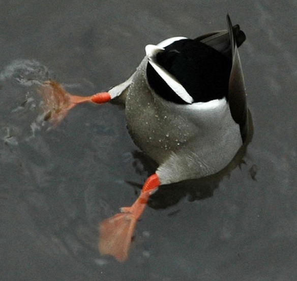
Ever wondered how a duck's rear end smells?

Ever wondered how a duck’s rear end smells?

So lets tease this study apart and look at the rationales and the results. First of all, influenza A virus infection in Mallards is a gastrointestinal infection, with viruses primarily replicating in the cells of the smaller intestine. Virus progeny is released in huge amounts with feces out into the environment. These well-known facts make the rear end a good starting point for studies on the possible olfactory difference between infected and uninfected birds. Mallards are fairly gregarious, although not extremely social birds, and the viruses released from the behind of one individual need to find its way to the front of the next individual. Mallards spend a large part of their lives dabbling – a behavior where surface water is taken into the mouth, the beak closes and the tongue presses upward, forcing the water through thin lamellae on the side of the beak. The yummy stuff, and perhaps viruses too, are stuck on the lamellae and is swallowed down. This behavior is thought to be one reason why Mallards (and other dabbling ducks) are especially frequent influenza A virus hosts.

Given this, it would actually be beneficial for a duck to be able to tell if the duck ‘over there’ is excreting viruses or not. Kimball et al. took this to heart and infected six domestic Mallards with an H5N2 low-pathogenic avian influenza virus. The researchers collected Mallard feces (the fancy word for bird shit) before experimental infection, and up till 10 days post infection. These fecal samples were then used to train mice – yes, you read it right: MICE – to differentiate between duck feces from infected and uninfected individuals. In the training sessions, mice were rewarded if they went to the right fecal sample (i.e. the infected one), and this was later tested in a double-blind test where both the mice and the operator had no clue where the different samples went in.

The tests showed that mice were actually quite good at learning to differentiate which birds that had been positive. The remainder of the paper examines which volatile compounds that were present in the feces and that may have given the results. For those of you that are keen chemists, I suggest you read the original article. For the rest of us, let’s just settle with that there were differences in the chemical spectra of the two groups. Particularly, acetoin (3-hydroxy-2-butanone) was more prevalent in the fecal samples from the infected birds.

Let us pause here and summarize. Mice can be trained to distinguish the smell of fecal samples from an influenza-infected duck. End of story. But a rather fun story. And not too far-fetched. The volatiles associated with infections are coming more and more in medicine, all the way from dogs trained at sniffing out cancers, to breath test to diagnose Helicobacter pylori infections. Smell is the future. However, the results of the current publication are not strong enough to say much on the use of olfactory cues among Mallards. Unfortunately, the paper is very thin in the material and methods section and the scant information on the infection protocol, the methods used for detecting influenza A virus in the samples, and for how long individual birds where shedding virus aren’t very helpful. If we are to believe the results we also need to be able to read all relevant information. For instance, the sex of each bird, whether control birds (if they indeed where controls) were housed together with the others, or separately, whether the diet and husbandry was the same for all birds etc. Many small questions, but where answers are important for interpretation.

And the bigger question, of course, is whether ducks themselves can distinguish the fragrance of infection. And whether that translates into a modified behavior. My personal feeling is that a duck with its bill spooning up water with muck and filth like a boss probably doesn’t take the time to smell the roses.

Don't forget to stop and smell the roses!

Don’t forget to stop and smell the roses!

Citation: Kimball BA, Yamazaki K, Kohler D, Bowen RA, Muth JP, et al. (2013) Avian Influenza Infection Alters Fecal Odor in Mallards. PLoS ONE 8(10): e75411. doi:10.1371/journal.pone.0075411

How bats in Peru change our view of flu (and it rhymes!)

By Jonas Waldenström

I am the real Bat Man and here to bite y'all

I am the real Bat Man and here to bite y’all

One of the major news in the virology community last year was the publication in PNAS describing a completely new influenza A virus. In line with the taxonomy traditionally used for influenza viruses it got the name H17N10, illustrating that it possessed novel hemagglutinin (H17) and neuraminidase variants (N10). However, it wasn’t the numbers that was the ground breaking news, it was the fact that the virus was detected in a Central American bat, and not in a bird. A tropical bat is very far from the ‘normal’ diversity of influenza A viruses seen in wetland birds and waterfowl. Although bats and ducks both have wings, in evolutionary terms they separate a very, very long time ago in the age of dinosaurs. In fact, there are more differences than similarities between bats and gulls in ecology, physiology and aspects of cellular biology. Hence, the bat flu was a remarkable observation. A real shaker. In one sweep, the whole flu field needed to come with terms that not all viruses are bird viruses.

The initial findings also hinted that the first bat influenza virus was unlikely to be alone. An influenza-iceberg, of sorts, made up of fluffy, winged mammals. This week, a first follow-up was published in PLOS Pathogens. A crew of (mainly American) scientists analyzed samples from bats sampled in the Amazonian parts of Peru in 2010, collected as part of CDC’s tropical pathogen surveys. In total, 114 individuals of 18 bat species were taken out from the freezers and different sample types were screened with a molecular method designed to broadly pick-up the RNA of any influenza A virus. They got one hit from a fecal sample in a single bat! A lucky shot at the Tivoli, given the low sample size. Prompted by this, the authors brought in the big machinery and sequenced the totality of the genetic material in the samples from this poor, long-dead bat and used bioinformatic tools to resolve the genome of the virus that had infected its intestines. When bit by bit was added it became clear that it was indeed a completely new influenza A virus, very different from avian viruses, and similar, but still distinctly different from the earlier H17N10 bat virus. And the name? H18N11 of course!

Please take a close look at the figure below. It shows the phylogenetic relationships of each of the influenza A virus’ eight RNA segments – in black are all ‘non-bat viruses’ and in red the two new bat viruses H17N10 and H18N11. For all the segments coding for ‘internal’ proteins, i.e. those involved in the polymerase machinery or the structural properties of the virus, you see that the two bat viruses are always found in a neat little red outgroup. This signals a long evolution away from other known influenza A viruses. It is a little prematurely to say exactly how long, but the branch lengths indicate that this happened a long time ago.

Phylogenetic trees for the 8 different IAV segements, see http://www.plospathogens.org/article/info%3Adoi%2F10.1371%2Fjournal.ppat.1003657

Phylogenetic trees for the 8 different IAV segements, see http://www.plospathogens.org/article/info%3Adoi%2F10.1371%2Fjournal.ppat.1003657

Now look at the hemagglutinin and the neuraminidase trees (HA and NA, respectively). The same pattern is repeated for the NA, but not the HA. In fact, the two novel hemagglutinins are nested within avian hemagglutinins. How can we interpret this? At first this doesn’t make any sense, but one has to remember that influenza viruses don’t evolve in the same way you or me, trees, shrimps or ferns do. Influenza viruses can reassort, meaning that if two viruses of different origin infect the same cell the different RNA segments can be put in new combinations in the resulting virions. Imagine two decks of cards being shuffled, one red and one black, and that each virion randomly consists of a draw of card from the combined shuffled deck, sometimes red, sometimes black, and sometimes mixed.  This is a rapid way in which new variants can arise, and a reason behind the genesis of pandemic flu in humans.

Returning to the bats, it seems that bat and avian viruses have met in a not too distant evolutionary past, and that a HA variant have sailed into the bat influenza gene pool. It will be interesting to see how the picture changes when more bat viruses are sequenced. Has there been one reassortment event, followed by drift and a subsequent separation into H17 and H18? Or, has there been many? Are there, perhaps, avian H17 and H18 to be found in South American birds? What about bats in North America, Europe, Africa and Asia?

One thing we can be sure of is that there are more viruses waiting to be detected and described. One sign of this comes from the current paper. The authors used the sequenced genomes to construct recombinant HA and NA molecules (using fancy virologist tricks) and used these to build assays (ELISAs) where bat sera could be screened for antibodies against the new HA and NA variants. Where the molecular screening yielded one positive bat, the serology approach found 55 of 110 bats showing signs of having been infected with flu earlier in life. This clearly indicates that influenza viruses are widespread in Peruvian bats, and likely in other parts of the world too. Moreover, they found cases of bats with antibodies to one of the recombinant HA or NA, but not to the other, suggesting that are more combinations of HA/NA to be found.

Finally, and perhaps the most interestingly of all results was that the hemagglutinin of bat influenza viruses does not to behave in the same way as avian hemagglutinins. When a virus is to infect a cell it needs the hemagglutinin protein to serve as a key, docking with a sialic acid receptor – the lock – on the cell. If the key and the lock don’t fit infection will not occur. For instance, a major division between human flu and avian flu is the preferred conformation of a galactose residue on the sialic acid receptors. This little difference makes it hard for avian viruses to infect humans, and vice versa. But with bat viruses it seems sialic acid receptors are not used at all! Instead bat HA uses an unknown receptor for cell entry. Holy Moses!

More to follow shortly, I suppose. Major obstacle at present is the lack of a culturing method for bat influenza viruses. Neither cell lines nor eggs have worked so far. Without the means to grow the virus it is very tricky to study it. But there are many clever virologists out there, so it is likely not too far away.

But I still prefer feathers to fur, and will stick with ducks.

Links to articles:

Tong S, Zhu X, Li Y, Shi M, Zhang J, et al. (2013) New World Bats Harbor Diverse Influenza A Viruses. PLoS Pathog 9(10): e1003657. doi:10.1371/journal.ppat.1003657

Tong S, Li Y, Rivailler P, Conrardy C, Castillo DA, et al. (2012) A distinct lineage of influenza A virus from bats. Proc Natl Acad Sci USA 109: 4269–4274.

Antarctica on the horizon

By Jonas Waldenström

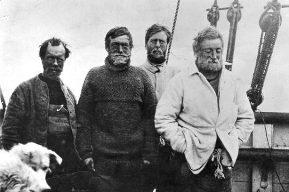
We had some great news last week! Our application to the Chilean research council was funded and our project Campylobacter in Antarctica can be launched in 2014! And what a launch it is: three years with fieldwork on the Antarctic Peninsula! Among penguins and skuas, whales and icebergs, seals and snow! Stuff for legends – Shackleton land, the last frontier – home of the brave!

The last frontier

The last frontier

The project is headed by Dr Daniel Gonzalez-Acuna from the University of Concepción in Chile – one of the best parasitologists in South America and a fantastic, enthusiastic guy! We have collaborated intensely for a number of years already, and last year was the closure of the first Antarctic program. That project aimed at understanding the life cycle of the seabird tick Ixodes uriae and its capacity of transmitting different diseases such as Borrelia, ornithosis, and others. Remember: this is a barren, dry and extremely cold environment, and it is incredible to think that this is also the preferred habitat for a parasitic arthropod. The penguin chicks – the provider of blood meals – are only available during the short Austral summer, and a life cycle can take many years to complete.

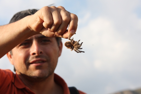
Dr Daniel Gonzalez-Acuna and an eight legged fellow

Dr Daniel Gonzalez-Acuna and an eight legged fellow

Most of the results of the previous expeditions are still in the lab, or are being analyzed in the computer. But some stuff is already out. During one of the expeditions a number of samples were collected from the sea just outside the scientific bases at different intervals from the bases’ sewage outlets. The water was filtrated and the filters cultivated for the presence of fecal coliform bacteria. In cases where bacterial growth was present, we went on and identified the bacteria and tested their susceptibility to different antibiotics. Much to our surprise we found real nasty bugs in the samples – ESBL-producing E. coli. The same type of bugs that can cause hospital-acquired infections in many parts of the world. Definitely not bugs to be leaked out into the Antarctic. This article, published in 2012 in AEM, was picked up by the press and spread around widely!

This time we will look at Campylobacter, especially Campylobacter jejuni, but also Campylobacter coli and Campylobacter lari. C. jejuni is a leading cause of bacterial gastroenteritis in humans. However, humans are only accidental hosts for this zoonotic pathogen. It has been detected from a wide range of animal species, primarily birds – and very frequently in poultry. This is likely where you, dear reader, may have made its acquaintance. Although the number of asymptomatic infections probably is high – some say very high – if you get bad, it is real bad. Stomach pains, vomiting and profuse diarrhea! All nasty, real nasty.

An army of locals on the march (photo by Jonas Bonnedahl)

An army of locals on the march (photo by Jonas Bonnedahl)

Why penguins? First, it is interesting to make a thorough inventory of what is in Antarctic wildlife. Are the campylobacters found in Antarctica similar to those in other areas of the world, and to disease-causing strains? However, equally interesting are the population genetic structure of campylobacters, and the frequent horizontal transmission of genes within and even between species. Like a jigsaw puzzle, where genome parts are exchanged and rearranged in new constellations. And now when all tools are available: which genes are found in strains adapted to live in cold environments? What do they do, and how do they work?

Many questions, and many good reasons to go to Antarctica. Time to pack my Shackleton outfit!

Proper researchers in field fittings

Proper researchers in field fittings

Hernández, J., Stedt, J., Bonnedahl, J., Molin, Y., Drobni, M., Calisto-Ulloa, N., Gomez-Fuentes, C., Soledad Astorga-España, M., González-Acuña, D., Waldenström, J., Blomqvist, M. & Olsen, B. 2012. Extended Spectrum β-Lactamase (ESBL), Antarctica. Applied and Environmental Microbiology 78: 2056-2058. [doi: 10.1128/AEM.07320-11]